7 common errors in FluoroSpot and how to avoid them
Published: December 13, 2023
Updated: June 11, 2025
7 minute read
Authored by: Tyler Sandberg
We’ve been doing FluoroSpot for quite some time and during all those years, we’ve seen FluoroSpot work great and, unfortunately, sometimes not so great. Luckily for you, we’ve already come across all the mistakes and have learned from them to make the best kits available. Here we’ll discuss 7 of the most common mistakes we’ve encountered in FluoroSpot and how to avoid them.
1. High background fluorescence
One of the most common issues our customers encounter is a high fluorescent background. There are several reasons why this may occur.

| Error | Solution |
|---|---|
| Cells weren't washed prior to addition to the FluoroSpot plate. | If the cells sit too long in the media before plating, cytokines or antibodies of interest could already be secreted in the media. If this media isn’t replaced before plating, the analyte of interest could be captured all over the plate, giving rise to high background. Simply washing the cells and resuspending them with fresh media before plating should lower background issues. |
| Reader exposure and contrast settings off. | In some readers, high exposure or improper contrast settings can give the appearance of high background. Adjust these and reread the plate. Or why not try IRIS 2, in which brightness and contrast can be adjusted after reading the plate? |
| Too many cells. | If a high proportion of cells respond in your wells, this can lead to a carpeting of cytokine or antibody in the wells. This may appear as high background when, in fact, it’s just confluent spots. Try reducing the cell number or incubation time. |
| Fluorescence enhancer not removed properly. | If excess Fluorescence enhancer is left in the plate, the wells can appear illuminated or give way to blurry spots. Properly decanting the plate against paper towels can help ensure the plates are completely empty. |
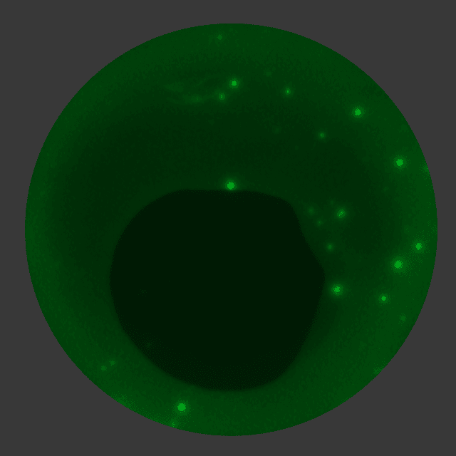
2. Glowing wells
Another regular concern we get from customers is a glowing center in FluoroSpot wells. After running the FluoroSpot assay, we all want to get right into data analysis as fast as possible. Our impatience often leads to reading a wet plate that gives rise to glowing centers. But fear not! Simply let the plate dry completely in the dark and then reread it.
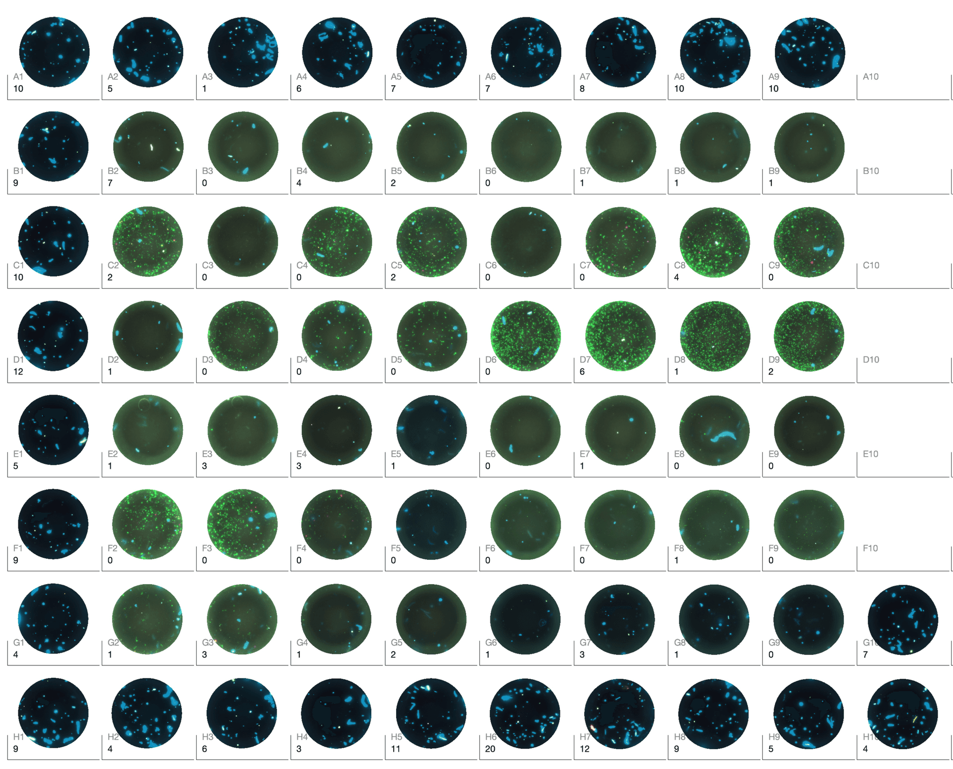
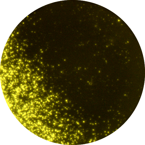
3. Spots only on the edges
Another pretty common error we see in FluoroSpot is an uneven distribution of spots in wells giving rise to a ring or crescent moon shape. This is almost always due to the addition of another reagent after cells were already added to the FluoroSpot wells. This addition “pushes” the cells in the plate to the edges causing them to settle there. This higher concentration of cells near the well edges can lead to confluent spots and negatively affect your spot counting. Yet another and similar issue is that the plates are not laid flat in the incubator causing the cells to pool to one side.
The solution here is extremely simple. Either always add your cells to wells last or mix cells with the stimulation cocktail before adding them to the plate. No more rings or moons! If you opt for the latter option, make sure to transfer the mixture to the FluoroSpot plate quickly, avoiding a build-up of analyte that could lead to a high background signal.

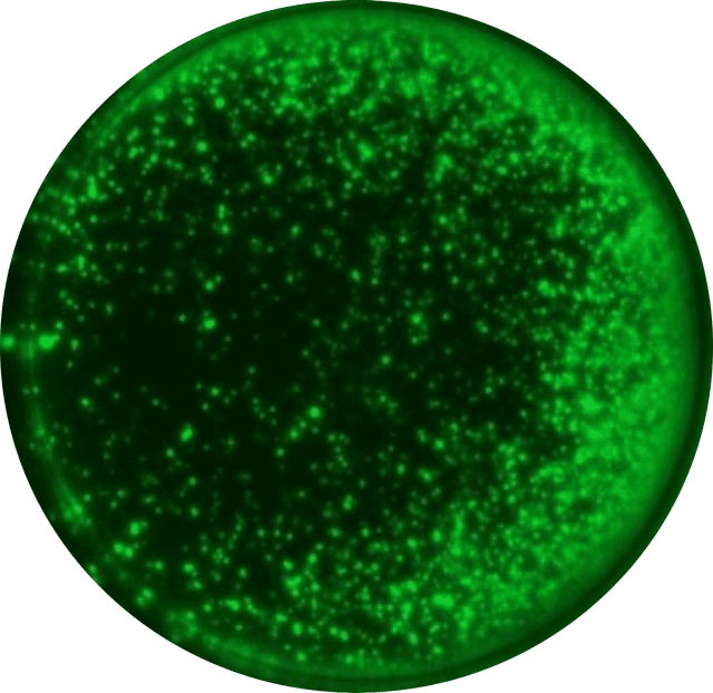

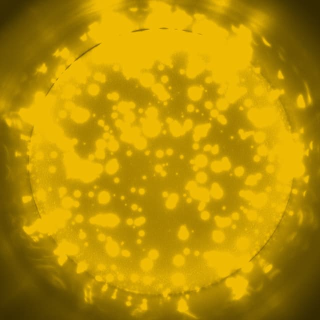
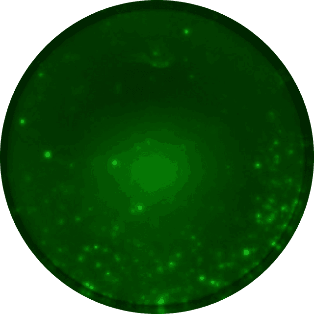
4. Capture effects
When capture antibodies with different specificities are coated together, the capture of one cytokine may affect the secretion of other downstream cytokines. This phenomenon is commonly referred to as capture effects or cytokine absorption effects. Capture effects can lead to very few or no spots at all.
For example, IL-2 capture antibodies may result in reduced activation of T cells, since capturing the secreted IL-2 decreases the amount of IL-2 available for downstream IL-2-dependent cytokine secretion. Luckily, capture effects can often be counteracted by adding an anti-CD28 mAb that provides a co-stimulatory signal to T cells, restoring cytokine secretion.

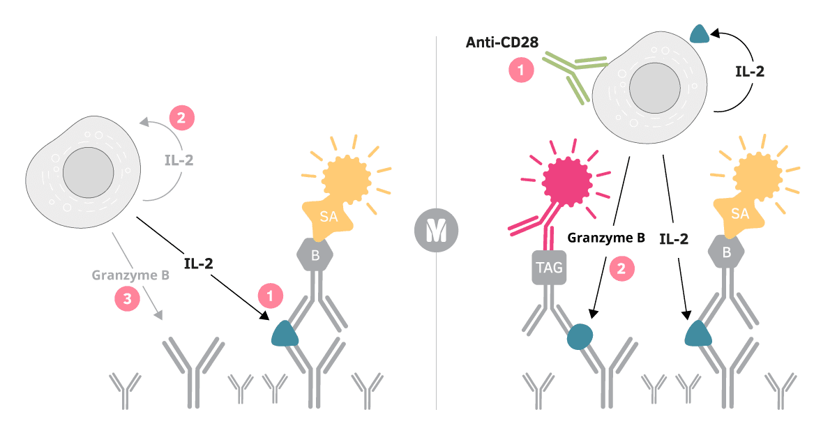
Capture effect (left image) 1) IL-2 secreted by the activated T cell is captured by coated anti-IL-2 capture antibodies. 2) As a result, IL-2-stimulation of the T cell itself (autocrine stimulation) as well as nearby T cells (paracrine stimulation) is impaired, ultimately leading to (3) decreased granzyme B secretion.
Counteraction (right image) 1) An anti-CD28 antibody can be added to provide a co-stimulatory signal that can (2) restore for example granzyme B responses.
5. No spots at all!
After reading your plate in a FluoroSpot reader, you see no spots in your wells. What happened? There could be several reasons why no spots show up in your FluoroSpot wells and here’s a table with the most common issues we’ve run into and how to solve them.
| Error | Solution |
|---|---|
| Low viability in the cell sample | Cells should have at least 89% viability to reliably detect antigen-specific responses. Try optimizing your isolation, freezing, and thawing protocols. |
| Suboptimal incubation time | Different analytes require different incubation times ranging from 12 to 72 hours. When running multiple analytes with FluoroSpot, always choose the incubation time for the slower analyte. Recommended incubation times can be found in the kit’s datasheet. |
| Reader exposure/contrast/camera settings | Some FluoroSpot readers require you to adjust exposure and contrast settings before reading the plate. These should be optimized prior to plate reading. And make sure external lamps are switched on in your reader. An easier solution would be to use the Mabtech IRIS 2 for all your FluoroSpot readings – its settings are already optimized for you. |
| Wrong fluorescent filter | All of our FluoroSpot kits work with the fluorescence filters fitted in IRIS and IRIS 2. Other FluoroSpot readers can have different filters, though, so make sure to consult your instrument’s specifications when purchasing our kits. |
| Bubbles in reagents | Ensure even distribution of cells and reagents when adding them to the wells. Bubbles can lead to areas with no spot formation. |
| Missed reagent | It can happen that a reagent or cell addition was missed in a single or multiple wells. Try a recovery protocol redoing the detection steps to see if spots can be detected. |
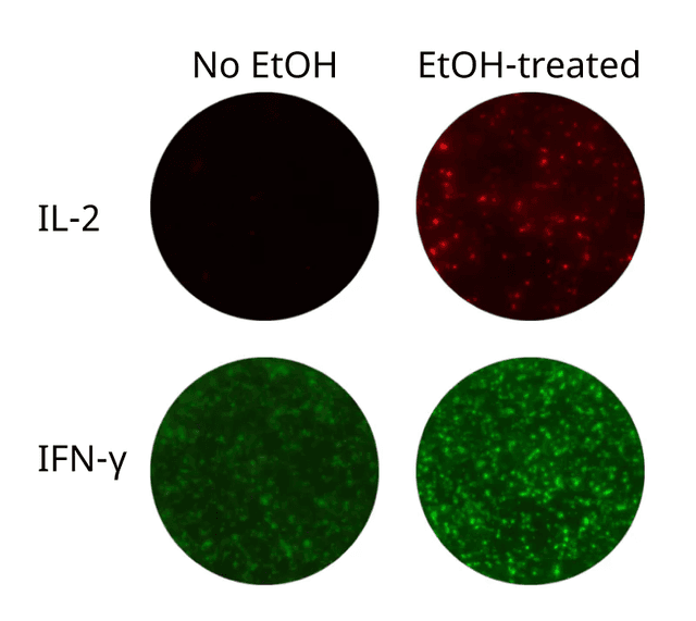
6. Improper ethanol treatment
Our FluoroSpot Flex kits allow you to customize your very own FluoroSpot combination and that means you coat the IPFL plates yourself. The PVDF membrane at the bottom of each well can only bind the required amount of capture antibodies after ethanol pre-treatment, which makes the membrane hydrophilic. Freshly prepared 35% ethanol solution should be used to activate the membrane for no more than 60 seconds.
The second issue is an uneven coating of the ethanol in the wells. This usually leads to a “bubbly” appearance, caused by no capture antibody being able to bind to the membrane. A simple solution is to tap the side of your plate after the addition of the ethanol to ensure even distribution over the membrane surface.
Don’t want to coat plates yourself? Check out our FluoroSpot Plus, Pro, and Path kits where the IPFL plates come pre-coated with the capture antibodies.

7. Lots of artifacts in my wells
Artifacts in FluoroSpot wells aren’t uncommon, especially when including the 380 (DAPI) channel in your readings. Small dust particles and hairs can get stuck to the membrane leading to artifacts in your wells. These are quite easy to remove with a masking tool, but it’s best to work to avoid them from the beginning. Here are some tips:
- Filtering reagents with a 0.2 µm sterile filter can remove dust and aggregates leading to cleaner wells.
- You can also try using a different type of paper towel or gauze for the final decanting step to reduce the amount of lint/dust exposure to the plates.
- Automated washers can also help reduce the number of artifacts with lower risks of particles and dust ending up in the wash