Anti-human IgG mAbs (MT78/145), biotin
Anti-human IgG mAbs (MT78/145), biotin
In stock
Delivery 4-9 business days
Shipping $0
Complementary products
Complementary products
Documents
Human IgG mAb guide
Learn how to combine Mabtech’s antibodies for human IgG ELISA and ELISpot. Adjust the setup to detect either total IgG or antigen-specific IgG. Different antibody combinations of monoclonal antibodies have been selected for the detection of the human IgG subclasses in ELISA and ELISpot. There is no cross-reactivity to human IgA1, IgA2, IgM, and IgE.
Application guide: ELISA
The human IgG ELISA can be adapted to detect antigen-specific IgG or the total IgG in serum or plasma samples.
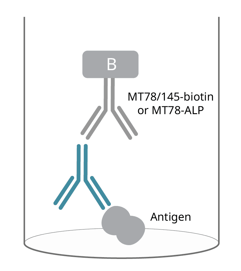
Antigen-specific IgG
Capture: Antigen
Detection mAbs: MT78/145, biotin or MT78, ALP
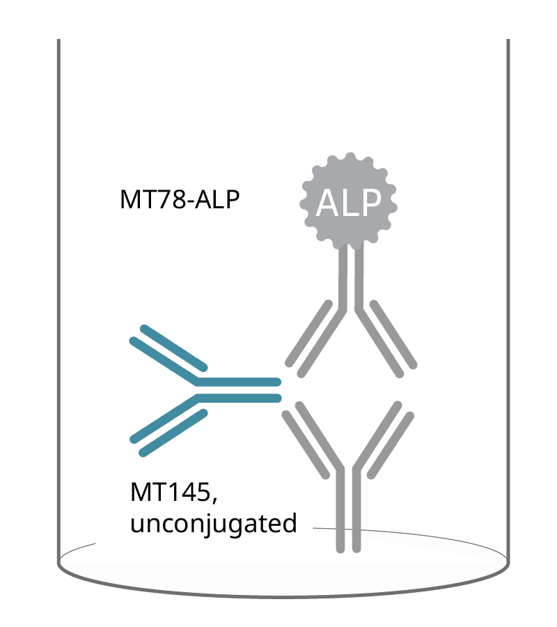
Total IgG
Capture mAb: MT145, unconjugated
Detection mAb: MT78, ALP
Application guide: ELISpot
The human IgG ELISpot and FluoroSpot can be adapted to detect antigen-specific IgG-secreting cells or the total IgG-secreting cells in cell samples like PBMCs.
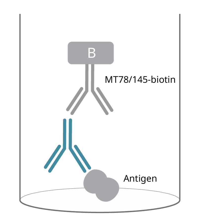
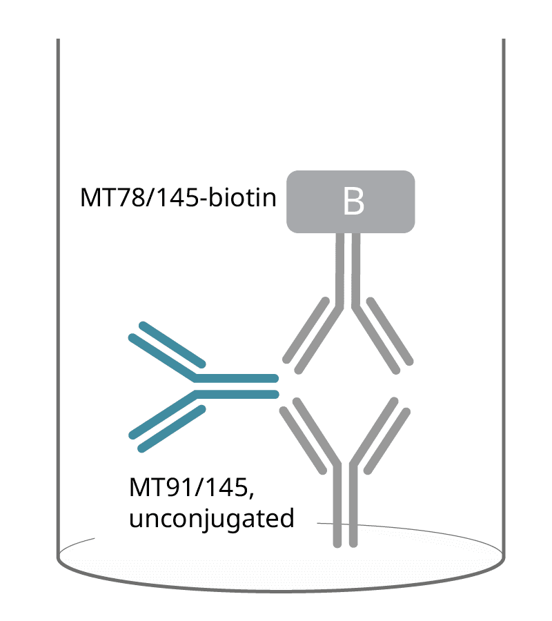
Antigen-specific IgG
Capture:
Antigen
Detection mAbs:
MT78/145, biotin
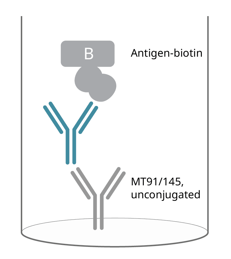
Capture mAbs:
MT91/145, unconjugated
Detection:
Antigen-biotin
Total IgG
Capture mAbs:
MT91/145, unconjugated
Detection mAbs:
MT78/145, biotin
Get inspired!
Make sure to check out how researchers have used Mabtech's human IgG ELISA, ELISpot, and FluoroSpot kits in everything from vaccine development to SARS-CoV-2 research. You can find it all in our Publication database!
Non-human primate cross-reactivity guide
The systems reactive with NHPs are either based on cross-reactive human kits or specifically developed monkey kits (NHP).
Cross-reactivity verification tests have been performed in ELISpot and/or ELISA by Mabtech and/or by others. Evaluations of assays with less solid evidence of cross-reactivity are shown as symbols within parentheses.
| ApoA1 | |||||||||
| ApoB | |||||||||
| ApoE (NHP) | |||||||||
| ApoE | |||||||||
| ApoH | |||||||||
| CCL2 (MCP-1) | |||||||||
| CCL4 (MIP-1β) | ( ) | ( ) | ( ) | ( ) | ( ) | ||||
| CCL22 (MDC) | ( ) | ( ) | ( ) | ( ) | ( ) | ||||
| CD25 | |||||||||
| GM-CSF | ( ) | ( ) | |||||||
| Granzyme A | |||||||||
| Granzyme B (NHP) | |||||||||
| Granzyme B | |||||||||
| IFN-α2 | |||||||||
| IFN-α pan | ( ) | ||||||||
| IFN-γ (NHP) | |||||||||
| IFN-γ | ( ) | ||||||||
| IgA (NHP) | |||||||||
| IgA | |||||||||
| IgG | |||||||||
| IgM | |||||||||
| INS | |||||||||
| IL-1α | |||||||||
| IL-1β | |||||||||
| IL-2 (NHP) | |||||||||
| IL-3 | |||||||||
| IL-4 | ( ) | ( ) | ( ) | ( ) | |||||
| IL-5 | ( ) | ( ) | ( ) | ||||||
| IL-6 | ( ) | ( ) | ( ) | ||||||
| IL-8 (NHP) | |||||||||
| IL-8 (ELISA) | |||||||||
| IL-8 (ELISpot) | |||||||||
| IL-10 (NHP) | |||||||||
| IL-10 | |||||||||
| IL-12/-23 (p40) | |||||||||
| IL-12 (p70) | |||||||||
| IL-13 (NHP) | ( ) | ( ) | ( ) | ||||||
| IL-13 | |||||||||
| IL-17A (NHP) | ( ) | ( ) | |||||||
| IL-17A | |||||||||
| IL-17F | |||||||||
| IL-17A/F | |||||||||
| IL-21 | |||||||||
| IL-22 | |||||||||
| IL-23 | |||||||||
| IL-27 | |||||||||
| IL-31 | |||||||||
| IP-10 | |||||||||
| Perforin | ( ) | ||||||||
| TGF-β1 (latent) | |||||||||
| TNF-α (NHP) | ( ) | ( ) | ( ) | ( ) | ( ) | ||||
| TNF-α | |||||||||
| CD3, mAb CD3-1 | |||||||||
| CD3, mAb CD3-2 | |||||||||
| CD28, mAb CD28-A | |||||||||
| IFN-γ mAb 1-D1K | ( ) | ( ) | ( ) | ( ) | ( ) | ( ) | |||
| IL-2 mAb MT8G10 | ( ) | ( ) | ( ) | ( ) | ( ) | ( ) | ( ) | ||
| IL-4 mAb IL4-3 | ( ) | ||||||||
| IL-17A mAb MT504 | |||||||||
| Perforin mAb Pf-344 | |||||||||
| TNF-α mAb MT15B15 | |||||||||
| IgG1 mAb MTG1218 | |||||||||
| IgG1 mAb MT1939 | |||||||||
| IgG2 mAb H6200 | |||||||||
| IgG2 mAb MTG211E | |||||||||
| IgG3 mAb MTG34 | |||||||||
| IgG4 mAb MTG42 | |||||||||
| IFN-γ mAb MT111W | ( ) | ( ) | ( ) | ( ) | ( ) | ( ) | ( ) | ( ) | ( ) |
| TNF-α mAb MT15B15 | ( ) | ( ) | ( ) | ( ) | ( ) | ( ) | ( ) |
Symbol keys:
Verified by Mabtech and/or by others. Tests performed in ELISpot and/or ELISA.
- good
- poor
- no
Based on reports from others or by analysis using recombinant proteins.
- ( ) good
- ( ) poor
- ( ) no
Specifically developed monkey kits are marked with (NHP).
Blank boxesRepresent cross-reactivities not evaluated.
* Comprises several species. Cross-reactivity may have to be verified on a species basis.
Product highlights
Publications (214)
Analyte information
IgG
| Analyte description | Immunoglobulin G (IgG) is the most abundant Ig isotype in serum, making up approximately 80% of all serum immunoglobulins. In humans, there are four subclasses of immunoglobulin G, with the highest serum concentrations of IgG1 followed by IgG2, IgG3, and IgG4. In mice, the IgG subclasses are defined as IgG1, IgG2a/c, IgG2b, and IgG3. The IgG molecule consists of two heavy and two light chains (κ or λ), resulting in a molecule with two arms for antigen binding. High levels of IgG antibodies are induced following the initial IgM response in a typical immune response to antigens. |
| Alternative names | Immunoglobulin G, IgG, IgG3 |
| Cell type | B cell |
| Gene ID | 3500, 3501, 3502 |
You may also like

