Anti-mouse IFN-γ mAb (R4-6A2), biotin
Anti-mouse IFN-γ mAb (R4-6A2), biotin
In stock
Delivery 4-9 business days
Shipping $0
Complementary products
Complementary products
Documents
Analyte combinations in FluoroSpot
Find out which analyte combinations we have evaluated in T cell FluoroSpot and which combinations are affected by capture effects or not.
What is a capture effect?
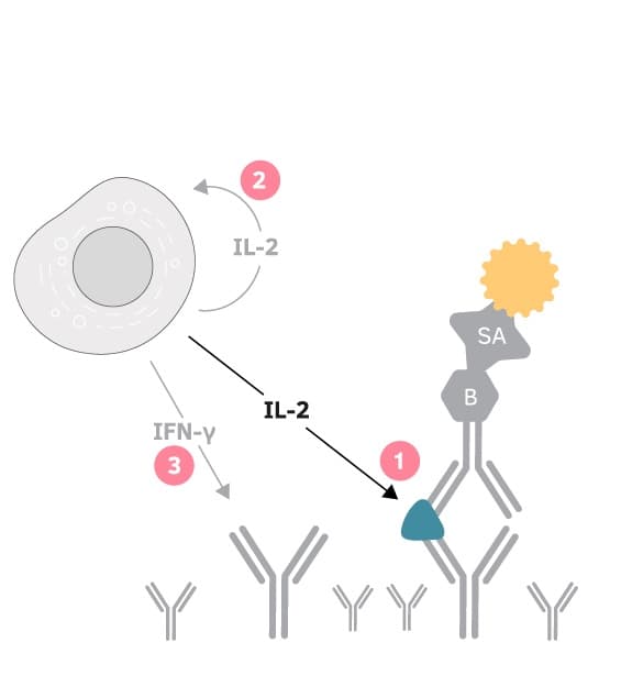
When capture antibodies with different specificities are coated together, the capture of one cytokine may affect the secretion of other cytokines. This is usually more pronounced when studying T cell responses with polyclonal stimuli compared to antigen-specific responses. Capture effects are seldom a problem and can often be counteracted by the addition of an anti-CD28 antibody.
(1) IL-2 secreted by the activated T cell is captured by coated anti-IL-2 capture antibodies. (2) As a result, IL-2-stimulation of the T cell itself (autocrine stimulation) as well as nearby T cells (paracrine stimulation) is impaired, ultimately leading to (3) decreased IFN-γ secretion.
Co-stimulation with anti-CD28
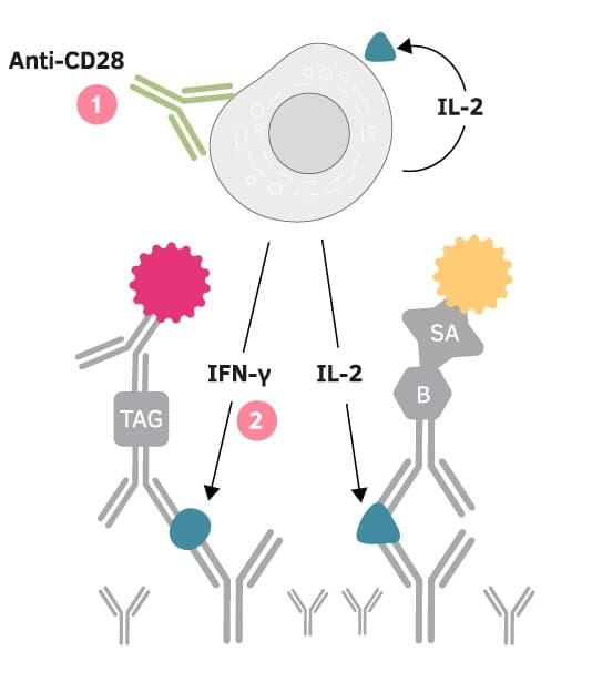
Anti-CD28 mAb provides a co-stimulatory signal to antigen-specific responses by binding to CD28 on T cells. The addition of an anti-CD28 mAb to the cell culture enhances antigen-specific responses and can counteract capture effects. For example, the presence of IL-2 capture antibodies may result in reduced activation of T cells, as capturing of IL-2 decreases the amount of available IL-2 and thereby dampens the IL-2-mediated stimulation of T cells. The addition of anti-CD28 mAb restores IFN-γ responses (depicted in the below images). Further optimization may be necessary, depending on the cells and stimuli used. Too high concentrations of the anti-CD28 mAb may result in an elevation of non-specific cytokine secretion.
(1) An anti-CD28 antibody can be added to provide a co-stimulatory signal that can restore (2) e.g. IFN-γ responses.
How to investigate capture effects
Our FluoroSpot Plus kits are evaluated for capture effects, and in studies of T cell responses we recommend co-stimulation with anti-CD28. With FluoroSpot Flex, it is possible to combine and build your own kit. For guidance look at our analyte combination table (above). Capture effects can be investigated by quantifying spot numbers in wells coated with single capture antibodies and compared to wells coated with a mixture of the capture antibodies. The compensatory effect of the anti-CD28 mAb may be assessed by comparing cells cultured with and without the anti-CD28 mAb.
Product highlights
Publications (1191)
Analyte information
IFN-γ
| Analyte description | Interferon-γ (IFN-γ) is the only type II interferon. This proinflammatory cytokine is secreted by activated T cells and NK cells. It activates macrophages and endothelial cells and regulates immune responses by affecting APCs, T cells, and B cells. Production of IFN-γ by helper T cells and cytotoxic T cells is a hallmark of the Th1-type phenotype. Thus, high-level production of IFN-γ is typically associated with effective host defense against intracellular pathogens. |
| Alternative names | Interferon-γ, Interferon-gamma, IFN-γ, IFN-gamma, IFN-g, IFNg, If2f, Ifg |
| Cell type | T cell, Tc, Th1, NK cell |
| Gene ID | 15978 |
You may also like


